Case Report
Maintaining the Sporting Shoulder after Injury; a Case Report on the Journey of Rehabilitation Exercise after Revision Rotator Cuff Repair
김은영1,†, 석순원2,†, 손종열2, 임재영3, 최중경4,*
회전근개 파열 재수술 후 스포츠재활의 성공적 여정: 증례보고
EunYoung Kim1,†, Soon Won Seok2,†, Jong Yeol Sohn2, Jae-Young Lim3, Joong Kyung Choi4,*
1Department of Rehabilitation Medicine, DM THE TNTN Clinic
2Department of Sports Rehabilitation, Yeonsung University
3Department of Rehabilitation Medicine, Seoul National University Bundang Hospital
4Department of Rehabilitation Medicine, St. Peter Covalescent Hospital, Siheung 15062, Korea
The first two authors contributed equally to this work.
*Corresponding Author: Joong Kyung Choi, M.D., Ph.D. Department of Rehabilitation Medicine, St. Peter Covalescent Hospital, 19, Pyeongansangga 5-gil, Siheung 15062, Korea, Tel: 82-10-9000-7353, Fax: 82-31-484-2800, E-mail:
cjk307@gmail.com
ⓒ Copyright 2023 Korean Academy of Sports Science and Exercise Medicine. This is an Open-Access article distributed under the terms of the Creative Commons Attribution Non-Commercial License (http://creativecommons.org/licenses/by-nc/4.0/) which permits unrestricted non-commercial use, distribution, and reproduction in any medium, provided the original work is properly cited.
Received: Feb 20, 2023; Accepted: Aug 18, 2023
Published Online: Aug 31, 2023
ABSTRACT
Surgical treatment of complete or full thickness tear of rotator cuff is a common treatment. However, the number of postoperative re-tear is increasing, and repetition of such surgical treatment causes various complications. Therefore, the importance of conservative rehabilitation exercise treatment has been emphasized, but there is no established rehabilitation protocol that shows a definite functional recovery of complete rotator cuff tear. The patient is a 46-year-old male patient who has already undergone an arthroscopic rotator cuff repair once due to a full thickness tear of the right supraspinous tendon. The patient visited the hospital after performing a second arthroscopic repair after diagnosing the re-tear of the supraspinous tendon. He visited the department of rehabilitation medicine and performed rehabilitation excise under physician supervision at cooperative rehabilitation center for 7 years, and showed complete functional recovery after rehabilitation treatment. The MRI without contrast performed 7 years after the surgery showed the maintenance of supraspinatus muscle and hypertrophy of surrounding muscles (especially deltoid muscle). And there is no functional limitation or pain. Therefore, functional recovery can be promoted through effective rehabilitation exercise protocol in complete or full thickness tear of rotator cuff, and even re-tear.
Keywords: Post-operative rehabilitation; Sporting shoulder; Rotator cuff tear; Rotator cuff repair surgery; Rehabilitation exercise; Retear after rotator cuff repair
Introduction
The number of rotator cuff tear patients is gradually increasing [1], and surgical repair is often performed first. However, due to the increase in the life expectancy of patients and the increase in sports activities, it is not uncommon to re-tear after surgery [2]. In particular, there is a limit to repairing again after re-tear, so conservative treatment or total arthroplasty is usually considered [3]. Therefore, if the rotator cuff is re-teared after one repair operation, rehabilitation exercise treatment for functional recovery would be a necessary treatment. However, the rehabilitation exercise protocol is being implemented in various ways in each hospital, and the rehabilitation program in a long-term way has not yet been established.
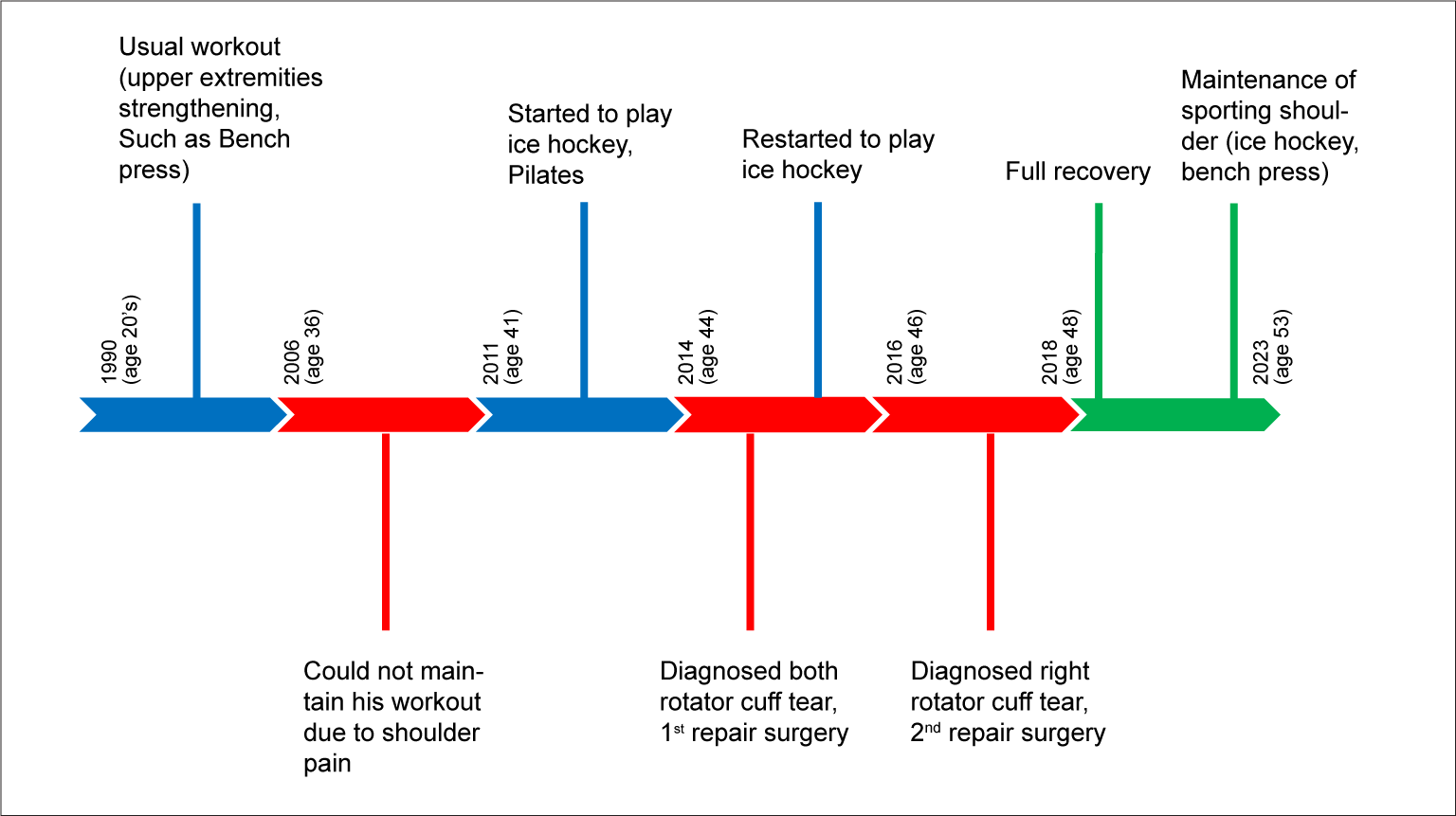
Therefore, we would like to present the rehabilitation journey during the 7-year follow-up of patient who underwent arthroscopic repair surgeries twice with two rotator cuff tears in the same area, and to illustrate the specific rehabilitation exercise protocol provided to the patient.
Case Report
A 46-year-old male patient underwent arthroscopic rotator cuff repair surgery on his right shoulder at an orthopedic clinic due to right shoulder pain that lasted for four months and visited one rehabilitation center. Two years before the visit, he had already undergone an arthroscopic repair once on the same shoulder due to full thickness tear of supraspinous tendon, and secondary repair was performed due to the findings of re-tear of the supraspinous tendon. There were symptoms that felt like touching while moving the arm and pain of NRS 5 before surgery.
He had been working out twice a week since his late 20’s. His workout consisted of mainly bench press or other upper extremity strengthening exercises. When he was 36 years old, he could not maintain his workout due to post exercise pain on both shoulder regions. Five years after that, he had started to play ice hockey and to do Pilates. He had played ice hockey for 2 hours a day and had done Pilates one hour per day twice a week before the first shoulder surgery. The Pilates program was both shoulder active resistive ROM exercise with Thera band for 40 minutes after 20 minutes massage around shoulder. One day he had experienced a big crash while playing ice hockey game. After that crash he could not move both shoulder and diagnosed with rotator cuff tear. At this time, after undergoing the first arthroscopic rotator cuff tear repair surgery on both sides, the pain improved and exercise was restarted. Two years later, after hitting his shoulder again, he was diagnosed with a right rotator cuff re-tear and underwent this second arthroscopic repair surgery (Fig. 1).
After second surgery both shoulder had 10 degrees of forward flexion, 15 degrees of abduction, less than 10 degrees of external and internal rotation in evaluation with a goniometer. The muscle strength of shoulders was grade 2 in all directions of movements. All passive range of motion of both shoulder could not be measured due to severe pain at that time.
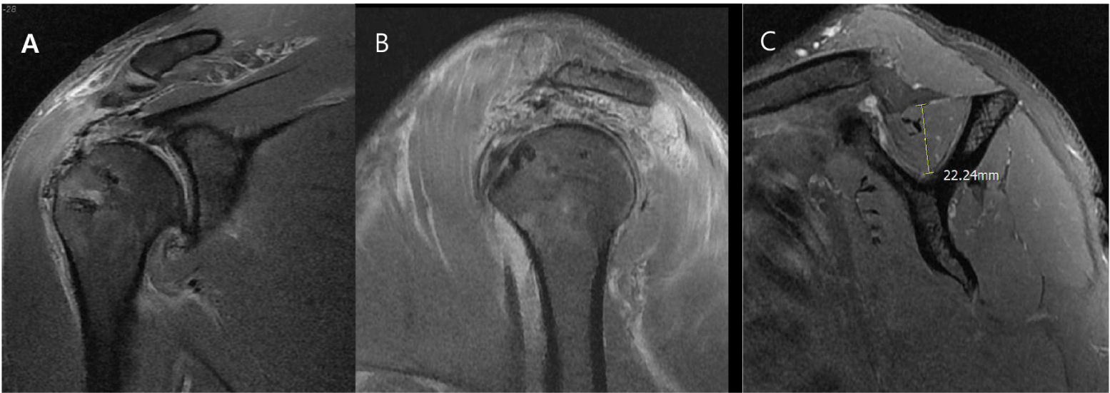
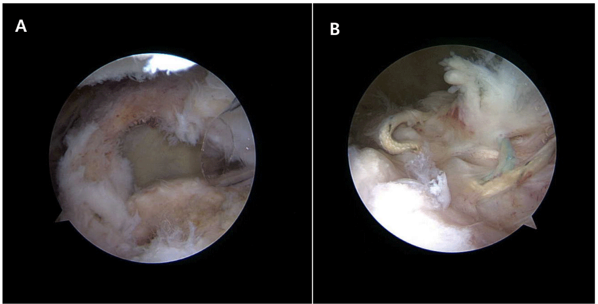
MRI performed after surgery showed that 2/1/0/0 grade atrophy of fat infiltration [4] of supraspinatus/infraspinatus/teres minor/subscapularis muscles, supraspinatus tendon was well attached in GT area, and another unusual post-op change was not seen (Fig. 2). In the arthroscopic findings, retraction of supraspinous tendon revealed exposure of it’s insertion site and loosening of the previous surgical suture materials (Fig. 3).
Fig. 2.
MRI of right shoulder after second repair, 2016’. A: Coronal plane view, B: Sagittal plane view, C: Sagittal plane view, Y view.
Download Original Figure
Fig. 3.
Arthroscopic findings within second surgery, 2016’. A: Re-tear lesion, B: Loosening of pre-inserted surgical materials.
Download Original Figure
Rehabilitation exercise program was under the physician’s supervision at cooperative rehabilitation center according to rehabilitation protocol 3 times a week (Table 1). Exercise was largely divided into three phases according to the painless shoulder abduction angle. Initially, not all phases and sets were completed, but the number and intensity could be gradually increased.
Table 1.
Rehabilitation Protocol*
|
1st Phase: |
Abduction AROM less than 30 degree
1. Body Weight Squat: 20 Repetitions, 3-5 Sets
Stand with both feet apart slightly wider than both hips. Place both hands on hips. Bend both knees till both thighs are nearly horizontal to floor. Keep upper body as straight as possible. Both knees were never in front of foot. Straighten legs back to standing position.
2. Incline Leg Press: 20 Repetitions, 3-5 Sets
Set up incline leg press machine. Both feet are in footplate with shoulder-width apart. Push the footplate away with heels and forefoot. Pause at the top of the movement. Do not lock out both knees. Return the footplate to the start position.
3. Leg Extension: 20 Repetitions, 3-5 Sets
Set up the leg extension machine and pad is in just proximal to ankle. Lift up weight till legs are almost straight. Hold hand bars and keep back against the backrest. Lower the weight back to starting position.
4. Leg Curl: 20 Repetitions, 3-5 Sets
Lie face down on leg curl machine. The rollers pad should rest just above the heels. Grasp the support handles on each side. Flex both knees till ankles are close to your buttock as possible. Return your feet to the starting position.
5. Side Lateral Raise: 20-30 Repetitions, 5 Sets
In standing position, Raise both arms with empty can position till pain free abduction range. Return arms to start position. Gradually weight up and increase abduction ROM. |
|
2nd Phase: |
Abduction AROM less than 90 degree
1. Smith Machine Incline Push Up: 10 Repetitions, 3 Sets
Set the Smith bar to xyphoid process level. Place hands on the bar, slightly wider than shoulder-width apart then step feet back so that body is in plank position. Make trunk 75 degree to the floor. Both arms should be perpendicular to your body. Lower down by flexing both elbows till chest nearly touched the bar. Return to the starting position by pressing through hands. Gradually lower the Smith bar level as muscle power increases.
2. Seated Cable Row: 20 Repetitions, 5 Sets
Sit on the bench and grasp the horizontal bar. Pull the weight up slightly off the stack, pull the bar into your upper abdomen. Slowly lower the weight to the starting position. Back must remain straight and keep lumbar lordosis. Do not let shoulders hunch over when arms are extended. Do not lean forward.
3. Lat Pull Down: 20 Repetitions, 5 Sets
Sit down on a pull-down machine with wide bar. Make sure that adjust the knee pad. Grab the bar with the palms. Extend both arms and bring the bar down until it touches upper chest. Raise the bar back to the starting position slowly when arms are fully extended and the lats are fully stretched. |
|
3rd Phase |
Abduction AROM more than 90 degree
1. Dumbbell Shoulder Shrug: 20-30 Repetitons, 5 Sets
In standing position, raise both shoulders as high as possible with dumbbell as if trying to touch them to ears. Gradually weight up and increase ROM
2. Overhead Cable Triceps Extension: 20-30 Repetitions, 5 Sets
Attach a rope to the bottom pulley of the pulley machine. Grasp the rope with both hands and turn away from the pulley such that back faces it. Bend body by placing one leg forward. Extend arms with hands directly above the head and both elbows should be close to head. Slowly lower the rope behind head as hold the upper arm stationary. Return to the start position by flexing triceps.
3. Dumbbell Pull Over: 20-30 Repetitions, 5 Sets
Hold a dumbbell with hands, roll back and lie on the bench. Extend arms toward the ceiling. Both palms should be facing each other, elbow slightly bent. Extend the dumbbell back and over the head slowly, not below the head. Return arms to the start position.
4. Incline Bench Press: 20-30 Repetitions, 5 Sets
Set up an incline bench to about 35-45 degree. Load the bar and lie back on the bench. Grasp the bar with slightly wider than shoulder-width. Both forearms are perpendicular to the floor. Pull the bar down with control to upper chest. Push the bar up and pull down.
5. Chest Press: 20-30 Repetitions, 5 Sets
Sit the chest press machine. Grasp the handle with a full grip and maintain a neutral wrist position with wrists in line with forearms. Push outward until both arms are fully extended. Pause briefly at full extension. Bend both elbows and return to the start position. |
Download Excel Table
In the first phase of the exercise program, it mainly focused on strengthening the muscles of the lower extremities and the trunk. After the exercise of the first phase was completely performed without pain, the second phase exercise was started. The second phase of exercise mainly focused on strengthening the large muscles behind the upper and lower back. The last three stages of exercise included small muscle exercises for the muscles that actually make up the shoulder girder and the rotor cuff. A patient had workout 3 times a week, every session was nearly one hour. After 2 years rehabilitation exercise, he finally achieved full recovery of right shoulder function.
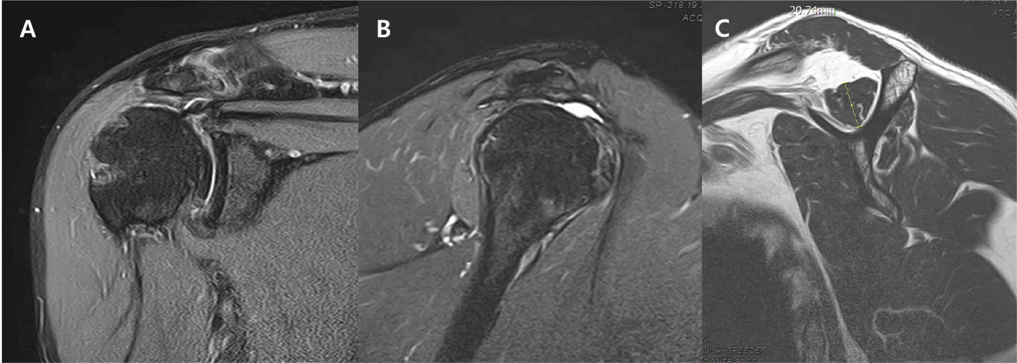
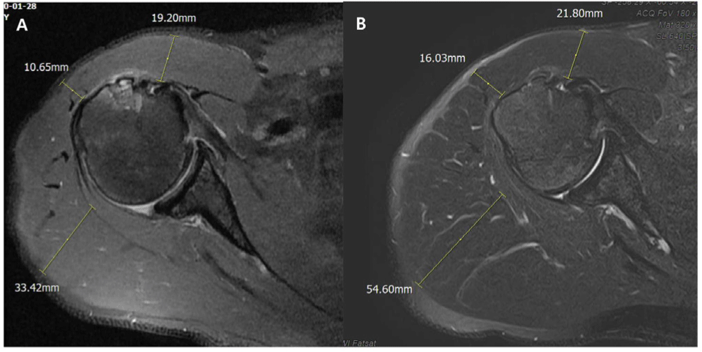
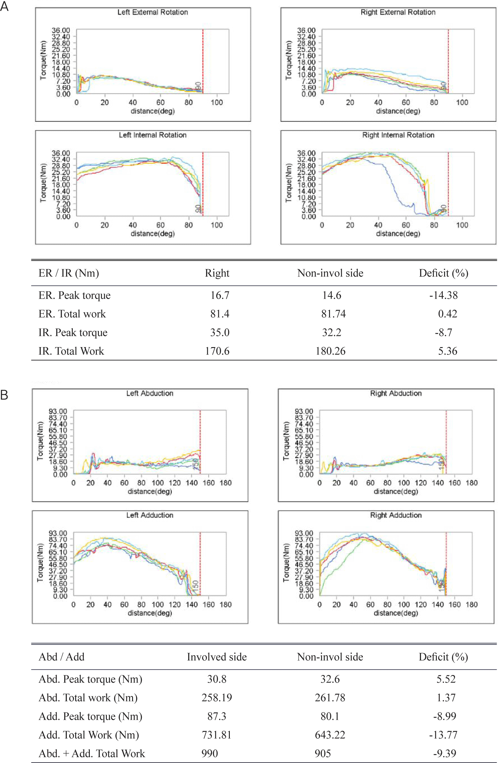
MRI performed after rehabilitation treatment showed a supraspinous tendon thinning but maintaining continuity, and another tear in the other cuff teandons was not reported, and 2/2/0/0 grade atrophy of fat infiltration of supraspinatus/infraspinatus/teres minor/subscapularis muscles (Fig. 4). This was confirmed to be 18 mm on both the left and right sides of the supraspinous muscle thickness measured by ultrasound (Fig. 5). Compared to before surgery, the thickness of the deltoid muscle increased from 19.2/10.7/33.4 mm to 21.8/16.0/54.6 mm in the anterior/middle/posterior muscles [5] (Fig. 6). Compared to the non-involved side in the isokinetic evaluation performed after rehabilitation treatment, there was no significant left-right difference in the peak torque and total work of the shoulder where the surgery was performed (Fig. 7).
Fig. 4.
MRI of right shoulder after rehabilitation protocol, 2023’. A: Coronal plane view, B: Sagittal plane view, C: Sagittal plane view, Y view.
Download Original Figure
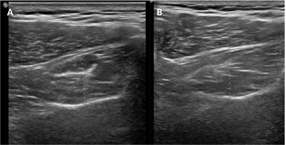
Fig. 5.
Sonographic findings of right shoulder after rehabilitation protocol, 2023’. A: Thickeness of right supraspinatus muscles, B: Thickeness of left supraspinatus muscles.
Download Original Figure
Fig. 6.
Differences in thickness of deltoid muscles over time. A: Before rehabilitation exercise, 2016’, B: After rehabilitation exercise, 2023’.
Download Original Figure
Fig. 7.
Isokinetic evaluation of both shoulder after rehabilitation exercise, 2023’. A: External and internal rotation of both side, B: Abduction and adduction of both side.
Download Original Figure
Discussion
The rotator cuff tear is very common, and there are many patients who have undergone the repair surgery accordingly. Damage to rotator cuff muscles at a young age occurs in highly active people, and therefore, there is a high possibility of further damage due to sports activities even after repair surgery [6,7].
In the case of the patient discussed above, he is also a patient with high sports activities, and after first surgery, a second surgery was performed due to a re-tear of the same area. Therefore, other treatment methods other than repeated surgery will have to be found.
Like this case, it is low at 65.9% in the case of maintaining the similar high sports level as before after the rotator cuff repair surgery [8]. However, for athletes following return to play criteria based on sufficient time and muscle strength, a high degree of the successful return and low retear rate is shown [9]. Therefore, rehabilitation exercise treatment through strengthening surrounding muscles could prevent not only additional muscular atrophy of the shoulder but also failure to return to high-level sports.
Patients after rotator cuff injury show changes in scapular position (protraction, depression, external rotation), and changes in cervical and thoracic curves (kyphotic posture). As the dyskinetic movement of the scapular is associated with shoulder pain [10], strengthening of the large posterior muscles of the spine and back should be prioritized for rehabilitation exercise treatment.
As shown in the above patient, stabilization of posterior muscles such as deltoid (especially posterior part) and teres minor plays a crucial role in the functional recovery of the shoulder as much as maintaining the muscle strength and integration of the supraspinatus muscle [8].
As an additional issue, the rehabilitation journey for this case has been successful enough to improve function and continue sports activities without further impairment and significant pain. Pataky et al. reported movement compensation driven by the deltoid muscles following severe rotator cuff tear contributed to produce a desired improvement in function [11]. However, since deltoid hypertrophy, right side was prominently confirmed in MRI, and a slight upward transfer of the humeral head was confirmed, the possible progression to rotator cuff arthropathy on a long-term standpoint should be considered. Through long-term follow-up observation, it is necessary to carefully balance the benefits obtained through enjoying sports performance sufficiently, and the harm caused by the secondary damage of the shoulder joint and the deterioration of joint function in the long run.